
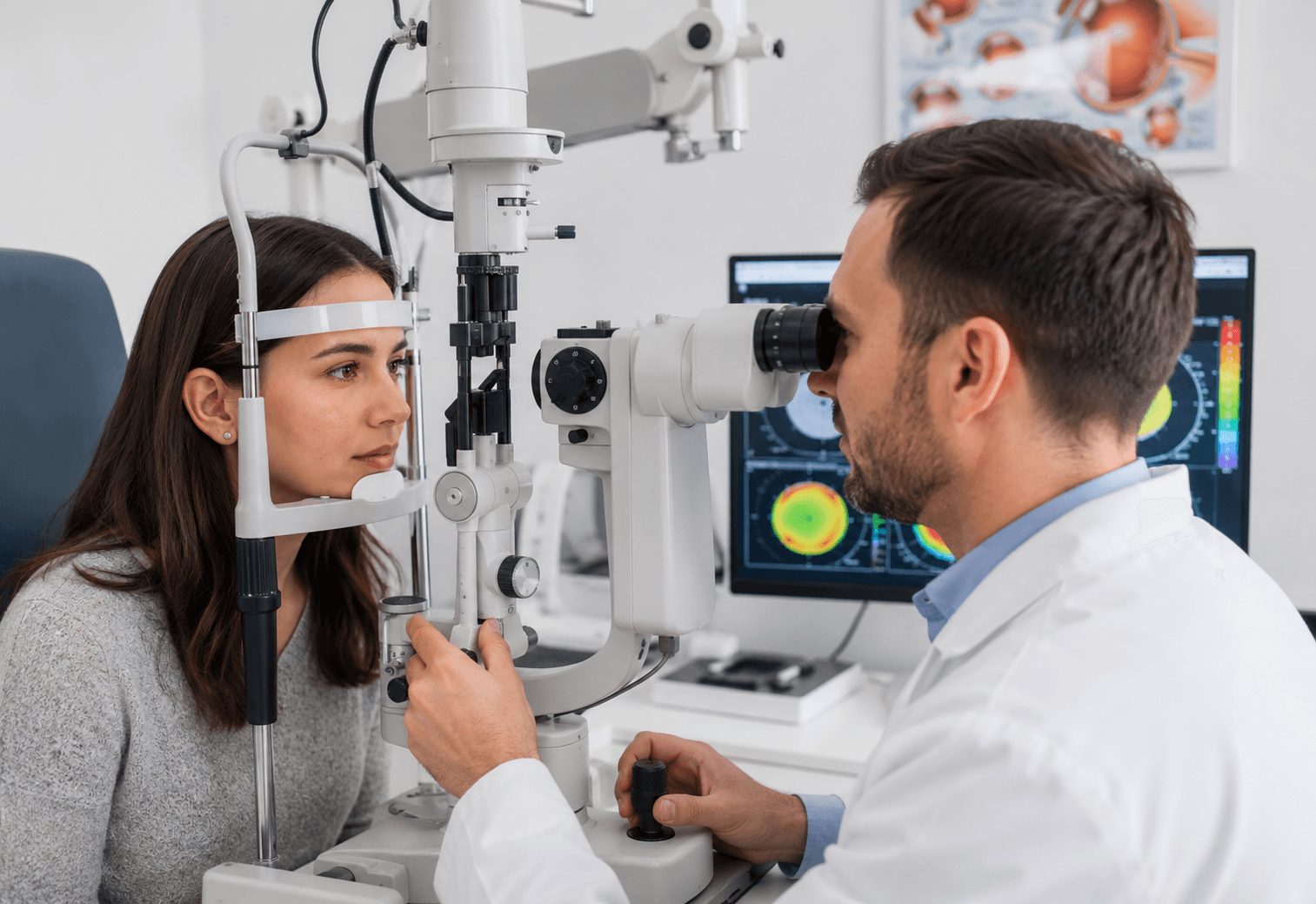
Doctors do not test vision as a single score. They measure each eye on its own, then measure how the two eyes perform together, and compare the results. That comparison is what reveals an imbalance that neither the patient nor the parent can detect at home.
A full assessment covers four things: how clearly each eye sees, how much focusing power each eye needs, whether the eyes are properly aligned, and how well they work as a team. Read together, these four measurements tell a doctor whether an eye simply needs a different prescription or whether amblyopia treatment is required. All of it forms part of understanding unequal vision and how it affects visual development.
Why the Difference Between Eyes Matters More Than Either Eye Alone
Two eyes that both test at a similar modest level are often perfectly normal that is simply a prescription. What draws clinical attention is a gap between them.
The concept that anchors this is best-corrected visual acuity: how clearly an eye can see after the right glasses are in place. An eye that is blurry without correction but sharp with it has an optical problem. An eye that stays blurry even with the correct lenses has a processing problem and that distinction determines everything that follows.
Step 1 Measuring Clarity in Each Eye Separately
Visual acuity testing
One eye is covered at a time so each is measured independently. Most people recognise the Snellen chart with its rows of shrinking letters; clinics increasingly use LogMAR charts, which have an equal number of letters per row and give a more precise comparison between the two eyes.
A result like 6/6 (or 20/20) describes what an eye can read at a standard distance compared with a normal reference. The number itself matters less than how the two eyes compare with each other.
Testing children who cannot read letters
Age is not a barrier to measurement. Young children are tested with picture charts or matching cards, where they point to a shape on a card rather than naming a letter. Infants and pre-verbal children can be assessed with preferential looking techniques, which use a child’s natural tendency to look towards a patterned image rather than a blank one.
Acuity is usually recorded both uncorrected and best-corrected. The difference between those two figures is one of the most informative numbers in the entire examination.
Step 2 Measuring Focusing Power (Refraction)
Objective refraction
Refraction determines the lens power each eye needs. Retinoscopy and autorefraction measure this objectively the examiner reads how light reflects from the back of the eye, so the result does not depend on the patient answering “better or worse.” This is essential for children and for anyone who cannot give reliable feedback.
Cycloplegic refraction in children
Children’s focusing muscles are strong enough to mask a genuine prescription, particularly for long-sightedness. Dilating drops temporarily relax that focusing so the true refractive error can be measured. The drops blur near vision and cause light sensitivity for a few hours, which is expected and temporary.
This step is what identifies anisometropia a meaningful difference in prescription between the two eyes, and one of the most common reasons one eye ends up seeing less clearly than the other.
Step 3 Checking Eye Alignment
Cover test and alternate cover test
The examiner covers one eye while watching the other. If the uncovered eye shifts to pick up fixation, it was not aiming at the target to begin with. In the alternate cover test, the cover moves back and forth between the eyes to reveal misalignment that stays hidden when both eyes are open.
Corneal light reflex and eye movement checks
A light held in front of the eyes should reflect from the same point on each cornea. An off-centre reflex suggests a turn. The examiner also tracks the eyes through a range of directions to check that both move smoothly and fully.
Together these checks show which eye the brain is actually fixating with — often the first clue that one eye is being favoured.
Step 4 Testing How the Two Eyes Work Together
Depth perception (stereopsis) testing
Stereopsis is the depth sense created when the brain fuses two slightly different views. It is tested with special 3D images viewed through filtered glasses, where the patient identifies which shape appears to stand out. The finest level of difference a person can detect is their stereoacuity a direct, measurable indicator of binocular teamwork.
Suppression testing
Separate checks reveal whether the brain is actively ignoring one eye’s input rather than combining it. If a patient consistently fails to see targets presented only to one eye while both are open, suppression is present. That is the mechanism explained in why the brain sometimes prefers one eye over the other.
This step is what separates a straightforward prescription difference from a genuine processing problem and it is the reason a full examination looks so different from a quick chart reading.
How Doctors Read the Results Together
No single measurement decides anything. The pattern across all four steps is what points to a diagnosis.
| What the tests show | What it typically indicates |
|---|---|
| Unequal prescription, both eyes correct to full acuity | An optical difference glasses or contact lenses are usually enough |
| Unequal prescription, one eye does not correct fully | Amblyopia suspected; treatment beyond correction is likely needed |
| Full acuity in both eyes but reduced stereopsis | A binocular coordination issue rather than a clarity issue |
| Misalignment with reduced acuity in the turned eye | A strabismic amblyopia pattern |
| Normal alignment, unequal acuity, unequal prescription | A refractive amblyopia pattern |
These are the patterns a clinician looks for, not a checklist for self-assessment. The same results can arise from other causes, which is why interpretation belongs with the examining doctor.
What Happens After the Measurements
Once the picture is clear, management follows a predictable order.
Correction comes first accurate glasses or contact lenses so both eyes receive the sharpest possible image. Any underlying obstruction or alignment problem is addressed next, since the brain will not change its habits while one eye is still handicapped.
Where suppression is confirmed and the weaker eye does not reach full acuity with correction alone, amblyopia treatment addresses how the brain processes that eye’s input. Binocular and dichoptic approaches present slightly different images to each eye so the task can only be completed by using both, encouraging fusion instead of suppression a different strategy from traditional patching, which blocks the stronger eye rather than training the pair.
Follow-up matters just as much as the initial diagnosis. The same measurements are repeated at intervals, so acuity gaps and stereoacuity become the yardstick for whether treatment is working.
When Should These Tests First Be Done?
Earlier examination gives a better result, because an imbalance detected before the brain has fully committed to one eye is easier to reverse. This is a large part of why early vision support matters during childhood.
It is also worth understanding the difference between a screening and a full examination. School and community screenings are quick checks designed to flag obvious problems, and they are valuable but they do not usually include refraction, alignment testing, or depth perception assessment. A child can pass a screening and still have a meaningful difference between the eyes. If any of the signs of unequal vision are present, a complete examination is the appropriate next step regardless of screening results.
Frequently Asked Questions
How do doctors test a child too young to read letters? Picture charts and matching cards are used for toddlers and preschoolers, while infants can be assessed with preferential looking techniques. Refraction is measured objectively, so no verbal response is required.
Why does my child need eye drops during the eye exam? Dilating drops relax the eye’s focusing muscles so the true prescription can be measured. Children’s focusing power is strong enough to hide a genuine refractive error without them. The blurring is temporary.
Can an eye test detect a lazy eye? Yes. Amblyopia is identified by comparing best-corrected visual acuity between the eyes alongside alignment and depth perception results. An eye that stays blurry with correct glasses is the defining finding.
What does it mean if one eye tests worse than the other? It may simply mean that eye needs a stronger prescription. It becomes more significant if the eye still underperforms after correction, which suggests the brain is not fully processing its input.
How often should these measurements be repeated? During active treatment, reassessment is typically frequent so progress can be tracked. Afterwards, routine intervals apply. Your eye care professional will set a schedule based on age and findings.